Are you a healthcare professional?

EOS-Inflammation-logo
No, I am not a healthcare professional
AstraZeneca Logo
  • Home
  • EOS Education
    • EOS Education
    • EOS Inflammation
    • SEA
    • EGPA
    • HES
  • Latest Science
  • Resource Library
  • EOS Education
  • SEA

SEA

Severe eosinophilic asthma (SEA) is the most common severe asthma phenotype, characterized by eosinophil-driven inflammation that leads to high exacerbation risk, decreased disease control, and progressive airway damage.1–3 Learn more about the central role of eosinophil-driven inflammation in severe asthma pathology, predictive biomarkers of disease, and treatment challenges below.

Module synopsis

The burden of eosinophil-driven inflammation in SEA

Eosinophils as drivers of disease pathology

Eosinophils as a reliable predictive biomarker

Impact of steroid management

Module synopsis

  • The majority of patients with severe asthma have an eosinophilic phenotype, and both blood and tissue eosinophilia are associated with pathophysiology in SEA1–3
  • As blood eosinophilia increases, the risk of severe exacerbations increases, disease control decreases, and lung function declines1–3
  • Eosinophils play a central role in asthma pathology, driving tissue dysfunction through the release of cytokines and multiple inflammatory mechanisms that cause airway damage and contribute to mucus plugging and airway wall thickening4,5
  • Blood and sputum eosinophil levels can predict future risk, making them reliable predictive biomarkers3,6–8
  • OCS are overused in patients with severe asthma, with their use associated with a substantial risk of adverse outcomes, including CV disease, metabolic disorders, and psychiatric conditions9,10
  • ICS remain a cornerstone of asthma management, but use of medium/high doses may increase the risk of side effects without additional benefit11

Back to top

The burden of eosinophil-driven inflammation in SEA

The majority of patients with severe asthma have an eosinophilic phenotype, with data from the ISAR estimating this as 83.8% of patients.2


In this study, patients most likely to have an eosinophilic phenotype (grade 3) were identified using the following clinical and biomarker features, as defined by an ISAR steering committee:2

  • Blood eosinophil counts ≥300 cells/µL
  • Receiving anti-IL-5 or anti-IL-5/R therapy
  • Blood eosinophil counts between 150 and 300 cells/µL and requiring maintenance OCS
  • Blood eosinophil counts between 150 and 300 cells/µL and at least two of the following: nasal polyps, elevated FeNO, or adult-onset disease

As blood eosinophilia increases, the risk of severe exacerbations increases, disease control decreases, and lung function declines, emphasizing the central role eosinophils play in asthma pathology.3,12


Eosinophilic inflammation in tissues is also recognized as a key contributor to the pathophysiology of several diseases, including SEA.13–15

Back to top

Eosinophils as drivers of disease pathology

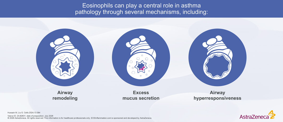
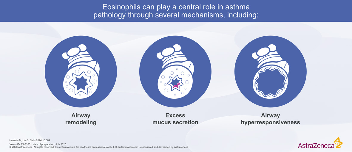
Role of Eosinophils in Asthma Pathology Role of Eosinophils in Asthma Pathology

Airway remodeling

Eosinophils play a central role in driving SEA pathology, through both direct and indirect mechanisms.12 Their activation is closely linked to the airway dysfunction seen in disease.12


When activated, eosinophils orchestrate tissue dysfunction through the release of a range of cytotoxic proteins which directly damage the airway epithelium.5,12,16–18 In parallel, eosinophils secrete pro-inflammatory cytokines and growth factors which promote fibroblast activation, extracellular matrix deposition, and airway wall thickening.16,19,20 Together, these pathological changes lead to progressive loss of epithelial integrity.19,20


Elevated eosinophil levels are associated with impaired structural integrity in asthma, with significantly greater epithelial damage observed in patients with high eosinophilic asthma than in those with lower eosinophil counts.20 In addition, sputum eosinophil levels have been positively correlated with basement membrane thickness, reinforcing the association between eosinophil-driven inflammation and structural airway change.19,20

Excess mucus secretion

Eosinophils also drive airway mucus pathology by increasing mucus plug size and stickiness through production of CLCs and EPX, which promotes oxidant formation and cross-linking of mucus.4,16,21 This makes mucus thicker and more elastic, making it harder to clear.4,22


Activated eosinophils in the airways undergo EETosis, which results in the formation of extracellular traps, release of alarmins that increase mucus viscosity and galectin-10, which results in the formation of CLCs.22


In patients with asthma, expression of MUC5AC by ​epithelial cells is increased, while expression of ​MUC5B is decreased. Reduced expression of MUC5B ​has been shown to promote eosinophil survival and ​is hypothesized to decrease mucus clearance, while ​MUC5AC increases mucus plugging. MUC5AC also plays roles in allergen-induced airway hyperresponsiveness and mucous metaplasia.23,24


Mucus plugs may persist in the same lung segment for years, and are associated with reduced lung function, airflow obstruction, frequent exacerbations, and increased mortality.4,25,26


Airway hyperresponsiveness

Eosinophils contribute to AHR through airway inflammation, structural remodeling, and functional changes in ASM.12 Eosinophil-derived mediators amplify type 2 inflammation, promoting epithelial injury and heightened bronchoconstrictor responses.12,27,28 Structural changes such as subepithelial fibrosis, basement membrane thickening, and increased ASM mass further reduce airway compliance and exacerbate AHR.16,20,28 In addition, eosinophils enhance ASM contractility and are associated with impaired bronchodilator responsiveness, sustaining airflow limitation and persistent symptoms in severe asthma.27–29


In addition to their direct roles in asthma pathology, eosinophils may also indirectly influence asthma pathology by promoting the activation, survival, ​and function of key effector cells, including T cells, B cells, neutrophils, and mast cells.5,16,17,30


For more information on the direct and indirect mechanisms of eosinophilic inflammation in pathophysiology, please visit the EOS inflammation module

Back to top

Eosinophils as a reliable predictive biomarker

The relationship between eosinophils and exacerbations / asthma control

Blood eosinophils are a reliable predictive biomarker of future risk and are an actionable target across patient subgroups.31


Monitoring blood eosinophils in patients with severe asthma has the potential to predict:

  • Severe exacerbations: higher eosinophil counts correlate with increased exacerbations3,6
  • Worse disease control: elevated eosinophils are associated with poorer symptom control1
  • Reduced lung function: higher eosinophil levels are also associated with significantly lower lung function including FEV1 below 50%, indicating more severe airflow limitation1,32

Blood eosinophil counts are a readily accessible and minimally invasive biomarker measurable from a simple peripheral blood sample.31 Routine monitoring of blood eosinophil count is straightforward in clinical practice and provides a practical way to assess eosinophil-driven inflammation, which may guide risk stratification and management decisions.2,31,33


Peripheral blood eosinophils have been shown to correlate with airway eosinophilic infiltration in sputum and appear to be a stronger predictor of severe exacerbation risk than total IgE, underscoring their value as a biomarker.33,34

A predict and prevent approach to disease management

There is some evidence to suggest that earlier intervention in SEA may be associated with a lower risk of:7,35,36

  • Severe airway exacerbations
  • Symptoms and morbidity
  • Lung function decline
  • Airway remodeling

Theoretically, earlier intervention may help to disrupt the inflammatory cascade before irreversible chronic structural damage within the airways develops or before the immunological cascade becomes self-perpetuating.7,37 In contrast, when treatment is delayed until the late or severe disease stage, it becomes more reactive, often referred to as a ‘firefighting’ approach.7 By this point, airway inflammation and remodeling may already be well established, making it harder to restore normal function.7,37


Some authors theorize that eosinophils may represent a prognostic marker that offers clinicians an opportunity for earlier intervention, supporting a predict and prevent approach that aims to reduce airway remodeling and preserve lung function in severe asthma.7,14

Back to top

Impact of steroid management

Corticosteroids have long been central to asthma management owing to their potent anti-inflammatory effects.38 ICS are recommended for maintenance therapy, while SCS (including OCS) have historically been used for the treatment of exacerbations and patients with severe or uncontrolled disease.9,11,38,39

OCS

OCS remain widely overused in asthma care.40,41


A retrospective, descriptive analysis of Canadian claims databases that included 88377 patients with severe asthma reported:

  • An average of 2.7 OCS claims per patient per year41
  • That 8.3% of patients with uncontrolled severe asthma had ten or more claims per year41

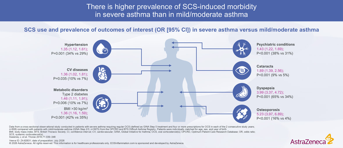
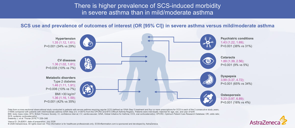
A cross-sectional observational study using UK primary care data that involved 808 patients with severe asthma and 3975 patients with mild or moderate asthma reported that:10

  • Patients with severe asthma typically have higher daily and cumulative doses compared with those with mild or moderate disease, reflecting the persistent inflammatory burden in this population10
  • This prolonged and repeated exposure to systemic steroid use places patients at substantial risk of adverse outcomes, including hypertension, CV disease, metabolic disorders such as type 2 diabetes, psychiatric conditions including anxiety and depression, cataracts, gastrointestinal complications such as dyspepsia, and osteoporosis10


SCS Induced Morbidity Severe Asthma Outcomes SCS Induced Morbidity Severe Asthma Outcomes


The widespread use of OCS highlights the need for steroid-sparing strategies that can control severe asthma while reducing the risk of corticosteroid
side effects.10,41

ICS

ICS remain a cornerstone of asthma management, with the potential to reduce symptoms, exacerbations, and asthma-related mortality.9,42 They are widely prescribed, with the majority of patients with persistent asthma receiving ICS therapy at some point during their disease course.43


Importantly, increasing ICS dose may not provide additional benefit.11 Recent estimates state that low-dose ICS (≤200 µg/day) achieves 80–90% of clinical benefit, while higher doses offer limited additional gain and are associated with an increased risk of clinically important side effects, including major CV events, arrhythmias, pulmonary embolism, and pneumonia.11,42


Reducing cumulative corticosteroid exposure, particularly OCS and, where appropriate, high-dose ICS, is an important therapeutic aim in asthma management, given the well-established risks associated with long-term steroid use.11,39,42,44

Back to top

AHR, airway hyperresponsiveness; ASM, airway smooth muscle; CLC, Charcot-Leyden crystal; CV, cardiovascular; EETosis, eosinophil extracellular trap cell death; EPX, eosinophil peroxidase; FeNO, fractional exhaled nitric oxide; FEV1, forced expiratory volume in 1 second; ICS, inhaled corticosteroid(s); IgE, immunoglobulin E; IL, interleukin; ISAR, International Severe Asthma Registry; MUC, mucin; OCS, oral corticosteroid(s); R, receptor; SCS, systemic corticosteroid(s); SEA, severe eosinophilic asthma

  1. Bumbacea D, et al. Eur Respir J 2004;24:122–128
  2. Heaney LG, et al. Chest 2021;160:814–830
  3. Jackson DJ, et al. Eur Respir Rev 2022;31:210150
  4. Dunican EM, et al. J Clin Invest 2018;128:997–1009
  5. Siddiqui S, et al. J Allergy Clin Immunol 2023;152:841–857
  6. Price DB, et al. Lancet Respir Med 2015;3:849–858
  7. Couillard S, et al. J Allergy Clin Immunol Pract 2023;11:704–712
  8. Schleich FN, et al. Respir Med X 2020;2:100022
  9. Bleecker ER, et al. World Allergy Organ J 2022;15:100726
  10. Sweeney J, et al. Thorax 2016;71:339–346
  11. Beasley R, et al. Am J Respir Crit Care Med 2019;199:1471–1477
  12. Hussain M, Liu G. Cells 2024;13:384
  13. Jacobsen EA, et al. Annu Rev Immunol 2021;39:719–757
  14. Quirce S, et al. Front Immunol 2023;14:1192284
  15. Klion AD, et al. Annu Rev Pathol 2020;15:179–209
  16. McBrien CN, Menzies-Gow A. Front Med (Lausanne) 2017;4:93
  17. Hogan SP, et al. Clin Exp Allergy 2008;38:709–750
  18. Arnold IC, Munitz A. Nat Rev Immunol 2024;24:858–877
  19. Broekema M, et al. Respir Med 2010;104:1254–1262
  20. Wilson SJ, et al. Clin Exp Allergy 2013;43:1342–1350
  21. Aegerter H, et al. Curr Opin Immunol 2021;72:72–78
  22. Ueki S, et al. Curr Allergy Asthma Rep 2019;19:35
  23. Lachowicz-Scroggins ME, et al. Am J Respir Crit Care Med 2016;194:1296–1299
  24. Venegas Garrido C, et al. Clin Exp Allergy 2022;52:1365–1378
  25. Chan R, et al. J Allergy Clin Immunol Pract 2023;11:195–199
  26. Venegas Garrido C, et al. Allergol Int 2024;73:351–361
  27. Janulaityte I, et al. Cells 2021;10:1389
  28. Bradding P, et al. J Allergy Clin Immunol 2024;153:1181–1193
  29. Jude JA, Panettieri RA Jr. Expert Rev Respir Med 2024;18:835–841
  30. Rosenberg HF, et al. Nat Rev Immunol 2013;13:9–22
  31. Fouka E, et al. Front Med (Lausanne) 2022;9:992565
  32. Bousquet J, et al. N Engl J Med 1990;323:1033–1039
  33. Zhang X-Y, et al. Clin Exp Allergy 2014;44:1137–1145
  34. Ban G-Y, et al. Allergy Asthma Immunol Res 2021;13:420–434
  35. Varricchi G, et al. Allergy 2022;77:3538–3552
  36. Thomas D, et al. Eur Respir J 2022;60:2102583
  37. Pavord ID, et al. J Allergy Clin Immunol Pract 2025;13:1574–1580
  38. Alangari AA. Ann Thorac Med 2014;9:187–192
  39. Volmer T, et al. Eur Respir J 2018;52:1800703
  40. Bleecker ER, et al. Am J Respir Crit Care Med 2020;201:276–293
  41. Côté A, et al. Ann Allergy Asthma Immunol 2025;135:403–411
  42. Bloom CI, et al. Am J Respir Crit Care Med 2025;211:54–63
  43. Bloom CI, et al. Eur Respir J 2019;53:1802130
  44. Sadatsafavi M, et al. Am J Respir Crit Care Med 2025;211:1165–1174
AZ-logo

EOSInflammation.com has been
developed by AstraZeneca.
For healthcare professionals only.
© 2026 AstraZeneca

  • Privacy Notice
  • Cookie Notice
  • Legal Statement
  • Contact Us

Veeva ID: Z4-82631
Date of preparation: July 2026