Are you a healthcare professional?

EOS-Inflammation-logo
No, I am not a healthcare professional
AstraZeneca Logo
  • Home
  • EOS Education
    • EOS Education
    • EOS Inflammation
    • SEA
    • EGPA
    • HES
  • Latest Science
  • Resource Library
  • EOS Education
  • HES

HES

HES (hypereosinophilic syndrome) is a heterogeneous group of rare disorders, often idiopathic, characterized by persistent blood and tissue hypereosinophilia, in which end-organ dysfunction and damage is mediated by eosinophilic infiltration and activation.1–4 Learn more about the role of eosinophilic inflammation in HES through this educational module.

Module synopsis

Defining HES and the critical role of eosinophilic inflammation

The diverse symptom burden of HES

The impact of management with corticosteroids and add-on immunosuppressive or cytotoxic therapies

Module synopsis

  • HES is a heterogeneous group of rare, often idiopathic, disorders characterized by blood and tissue hypereosinophilia1–4
  • A diagnosis of HES requires the presence of peripheral blood and tissue hypereosinophilia, evidence of eosinophil-induced organ damage and/or dysfunction, and the exclusion of other potential causes as the primary reason of organ damage3
  • The clinical manifestations of HES are variable and depend on the specific organs affected by eosinophilic infiltration5
  • End-organ damage and dysfunction due to eosinophilic infiltration involve several mechanisms, including tissue remodeling and fibrosis, as well as the release of various cationic proteins, cytokines, and lipid mediators from eosinophils with cytotoxic and/or procoagulant effects4 
  • Despite OCS and add-on immunosuppressive therapies, patients with HES continue to experience substantial disease burden, compounded by treatment-related toxicity6,7

Back to top

Defining HES and the critical role of eosinophilic inflammation

HES is a heterogeneous group of rare disorders characterized by persistent hypereosinophilia.2–4 Hypereosinophilia is defined as an absolute eosinophil count ≥1500 cells/µL in the peripheral blood on two examinations with an interval of ≥2 weeks.3 Tissue hypereosinophilia occurs when the percentage of eosinophils in the bone marrow section exceeds 20% of all nucleated cells, and/or pathologist-confirmed extensive eosinophil tissue infiltration is present, and/or marked deposition of eosinophil granule proteins is found (with or without eosinophil tissue infiltration).3

HES is diagnosed based on the presence of blood and tissue hypereosinophilia and evidence of eosinophil-induced organ damage, including tissue eosinophil infiltration and/or deposition of eosinophil-derived proteins (eg eMBP1 or EPX), supported by clinical, histopathological, and laboratory findings.3,4 For a diagnosis of HES to be established, it is important to exclude other disorders or conditions as a major reason for organ damage.3,4


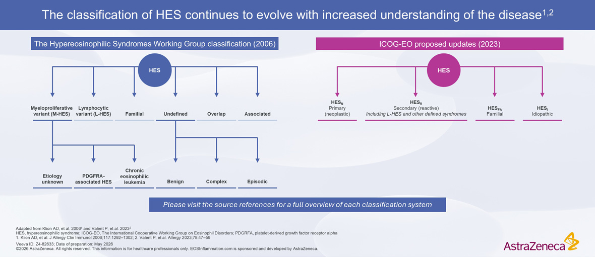
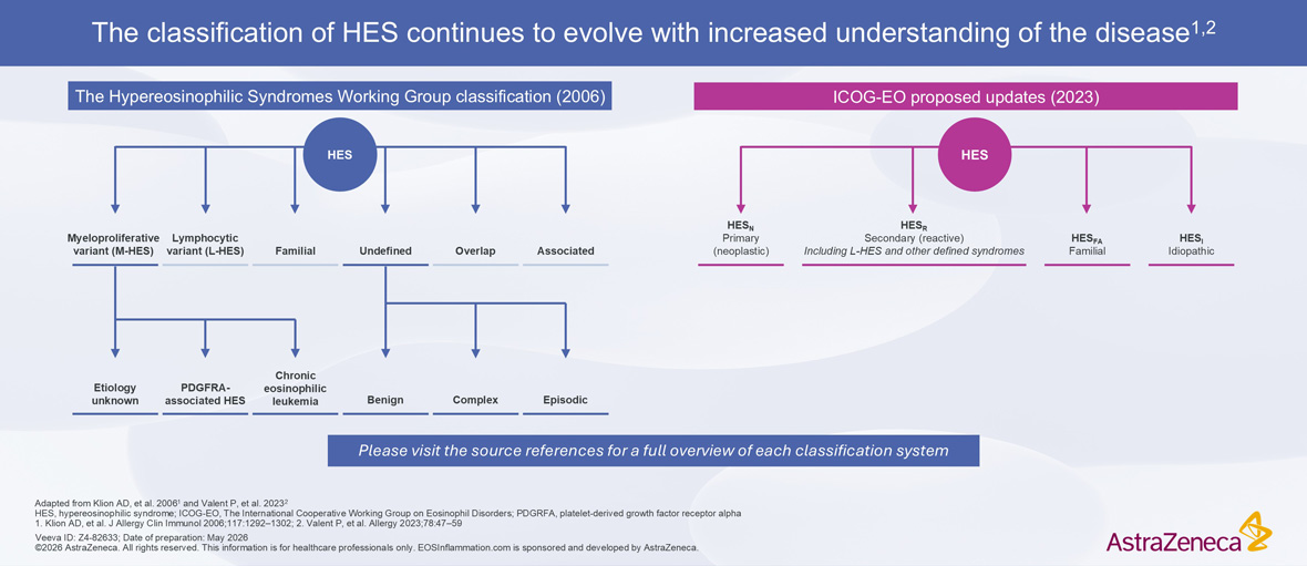
HES Classification Evolution From 2006 To 2023 HES Classification Evolution From 2006 To 2023


There are several clinical subtypes of HES, of which the classification continues to evolve with increasing understanding of the disease.3 In 2006, Klion et al. proposed a HES classification algorithm based on clinical phenotype.8 This included subtypes such as M-HES, characterized by documented or presumed clonal eosinophilic involvement (including the presence of the FIP1L1-PDGFRA fusion gene), and L-HES, defined by a clonal or phenotypically aberrant T cell population, among others.8 These classification categories remain widely used in clinical practice; however, updated recommendations now define HES based on underlying etiology.3 The updated categories are primary (neoplastic) HES, involving an underlying stem cell, myeloid, or eosinophil neoplasm; secondary (reactive) HES, involving an underlying condition in which eosinophils are considered non-clonal cells and hypereosinophilia is considered to be cytokine-driven; as well as familial (hereditary) or idiopathic (I-HES) forms.3 The etiology of HES is considered idiopathic in over 50% of patients, ie no underlying neoplastic or reactive condition is identified.1,9

Back to top

The diverse symptom burden of HES

Organ system involvement in HES is highly variable, resulting in a diverse symptom burden that differs between individuals and reflects eosinophil infiltration of affected organs.3,7


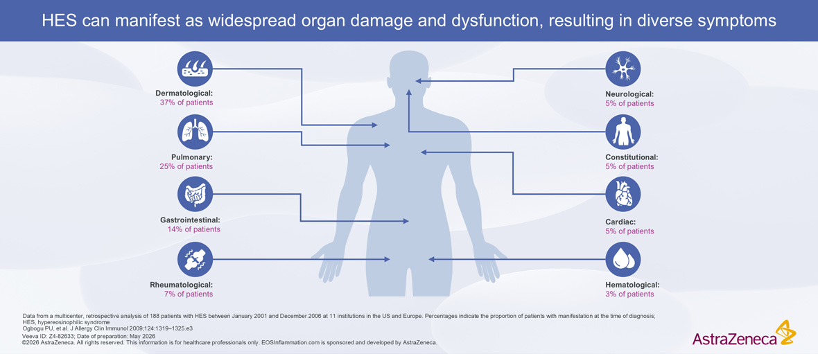
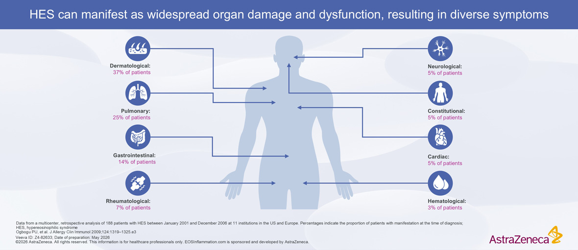
HES Clinical Manifestations and Organ Dysfunction Symptom Percentages HES Clinical Manifestations and Organ Dysfunction Symptom Percentages


In a multicenter, retrospective analysis of 188 patients with HES, the most common manifestations at initial presentation were dermatological, pulmonary, and gastrointestinal.7


Similarly, a more recent analysis of a US-based claims database of 212 patients with HES reported the most common disease manifestations to be those related to the upper airway/pulmonary (62%), constitutional (46%), dermatological (36%), gastrointestinal (34%), hematological (28%), and cardiovascular systems (24%).10


In a survey of 26 patients with HES, the most commonly reported symptoms were fatigue (65%), itch (50%), and shortness of breath (38%).5 Patients described fatigue as a “full body” sensation and compared it to “having the flu” or being “run over by a truck”.5


HES may affect multiple organ systems. Data from a real-world retrospective, observational US-based cohort study of 53 patients with I-HES or L-HES showed that 53% at baseline and 63% after one year of follow up had signs and symptoms of HES related to at least three different organ systems.11 Similarly, a retrospective, observational study of 79 patients at a single center in Italy reported that 80% had involvement of at least two organ systems, most commonly the lungs, skin, bowel, and the peripheral nervous system.12


Eosinophilic infiltration as a driver of end-organ damage and dysfunction in HES

End-organ damage and dysfunction in patients with HES due to eosinophilic infiltration occurs through several mechanisms.4,13,14 Eosinophils contain granules which can selectively release a range of cationic proteins, cytokines, (including IL-3, IL-5, IL-6, IL-13, TNF-α, and TGF-β), and lipid mediators that have a range of actions in tissues, including:

  • Cytotoxic effects4,13
  • Procoagulant effects4,13
  • Profibrogenic properties4,13,14

End-organ damage through tissue remodeling and fibrosis may partly explain the fibrotic lesions that can affect the skin and cardiac tissue in patients
with HES.4,15


For more detail on the role of eosinophils in pathology, visit the EOS inflammation module

Back to top

The impact of management with corticosteroids and add-on immunosuppressive or cytotoxic therapies

Management of HES has historically relied on the long-term or repeated use of OCS, with immunosuppressive or cytotoxic therapies used as add-on therapy to improve disease control in cases of inadequate response or as a CS-sparing strategy.9 Immunosuppressive or cytotoxic agents may also be associated with considerable toxicity.7


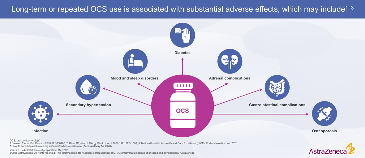
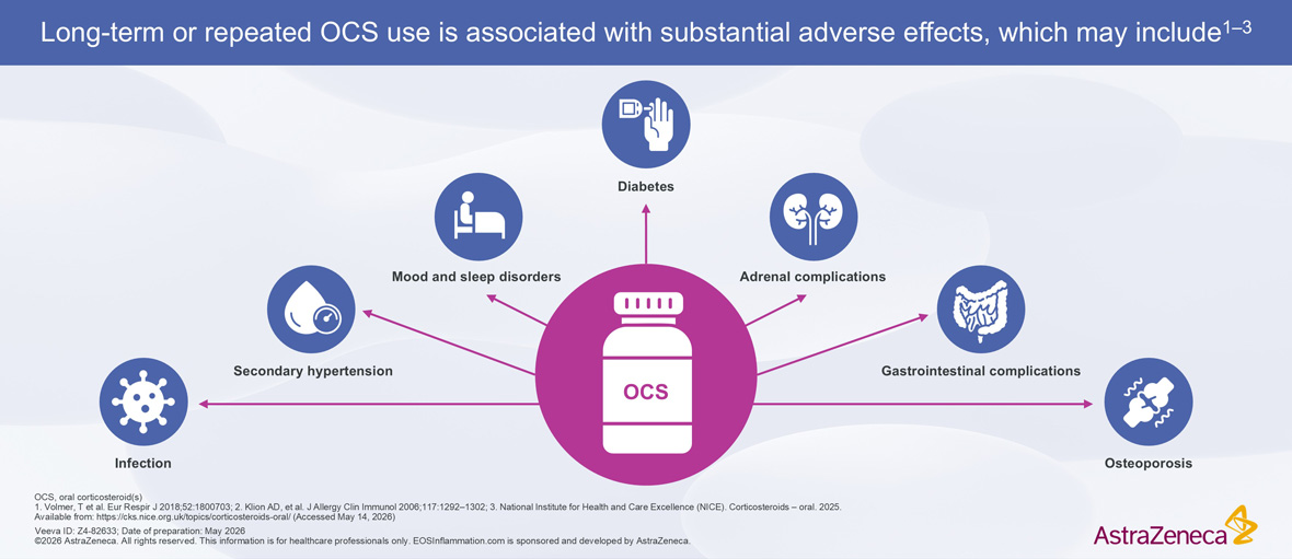
Adverse Effects Associated with Long-Term or Repeated OCS Treatment Adverse Effects Associated with Long-Term or Repeated OCS Treatment


In a retrospective, non-interventional medical chart review of 280 patients with HES in five European countries, use of OCS ranged from 80.3% of patients in France to 100% of patients in Germany, with long median durations of treatment (ranging from 8.5 to 23.1 months across countries) and high maximum daily doses (ranging from 24.8 to 42.4 mg/day).9


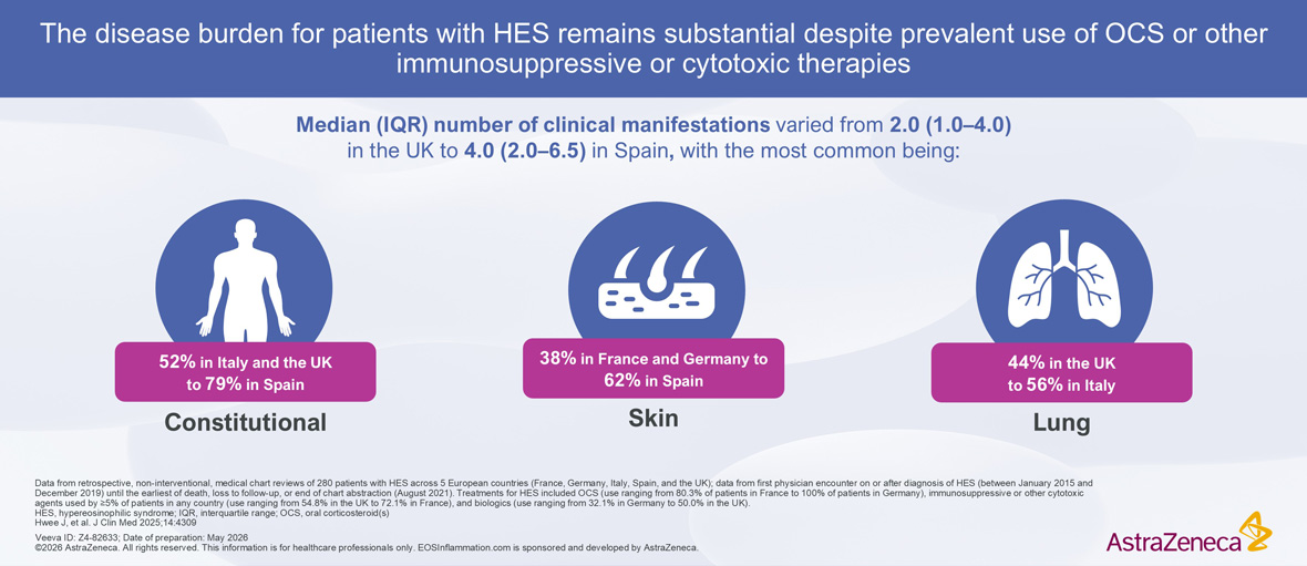
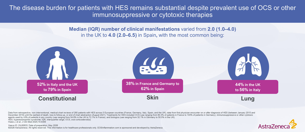
Immunosuppressive or cytotoxic agents were also widely used in this population (64% of patients), with imatinib (20%), azathioprine (14%), and methotrexate (10%) being the most commonly prescribed.9 According to this study, despite the extensive use of OCS and add-on therapies, a substantial proportion of patients (60%) did not achieve a complete clinical response (defined as a physician-reported improvement or resolution of symptoms, with a blood eosinophil count of ≤500 cells/µL).9 The disease burden was high, with the median (IQR) number of clinical manifestations varying from 2.0 (1.0–4.0) in the UK to 4.0 (2.0–6.5) in Spain. The most common were constitutional, lung, and skin involvement. The proportion of patients requiring hospitalization was highest in the UK (27/62, 43.5%) and lowest in Spain (6/52, 11.5%).9


Another multicenter, retrospective analysis of 188 patients with HES highlighted the lack of efficacy and high toxicity of second-line therapies, with the majority of patients discontinuing treatment with agents such as hydroxyurea, interferon-α, cyclosporin, and imatinib due to poor efficacy or intolerance.7


HES Clinical Manifestations and Disease Burden Despite OCS Therapies HES Clinical Manifestations and Disease Burden Despite OCS Therapies


Patients may also require higher CS doses to achieve a response (defined as the reduction of absolute eosinophil count to <1000/mm3 and control of symptoms).16 In a retrospective assessment of 164 patients with PDGFRA-negative HES, 91% of patients successfully responded to CS treatment. However:16

  • The majority of patients (61%) failed to respond to lower doses of CS (≤10 mg prednisone equivalent)
  • 52% of patients had a variable response to CS
  • 9% of patients had no response to CS
  • 19% of all CS responders (n=28) required a higher dose of ≥21 mg/day prednisone equivalent to achieve disease control

The toxicity associated with both CS and immunosuppressive therapies, combined with variable efficacy, a need for higher CS doses in many patients, and the persisting high disease burden, highlights a need for alternative approaches to the management of HES9,16

Back to top

CS, corticosteroid(s); eMBP1, eosinophil major basic protein; EPX, eosinophil peroxidase; FIP1L1, factor interacting with PAPOLA and CPSF1; HES, hypereosinophilic syndrome; I-HES, idiopathic HES; IL, interleukin; IQR, interquartile range; L-HES, lymphocytic HES; M-HES, myeloproliferative HES; OCS, oral corticosteroid(s); PDGFR, platelet-derived growth factor; TGF, transforming growth factor; TNF, tumor necrosis factor.

  1. Klion AD. Blood 2015;126:1069–1077
  2. Chen MM, et al. J Allergy Clin Immunol Pract 2022;10:1217–1228.e3
  3. Valent P, et al. Allergy 2023;78:47–59
  4. Groh M, et al. Orphanet J Rare Dis 2023;18:100
  5. Kovacs N, et al. J Allergy Clin Immunol Pract 2020;8:3209–3212
  6. Hwee J, et al. Ann Allergy Asthma Immunol 2023;130:768–775
  7. Ogbogu PU, et al. J Allergy Clin Immunol 2009;124:1319–1325.e3
  8. Klion AD, et al. J Allergy Clin Immunol 2006;117:1292–1302
  9. Hwee J, et al. J Clin Med 2025;14:4309
  10. Ogbogu PU, et al. J Allergy Clin Immunol Pract 2026;14:205–214
  11. Dolin P, et al. Blood 2024;144(Suppl. 1):2316–2317 (Abstract)
  12. Nicola S, et al. Front Immunol 2025;16:1735131
  13. Akuthota P, Weller PF. Immunol Allergy Clin North Am 2015;35:403–411
  14. Klion AD, et al. Annu Rev Pathol 2020;15:179–209
  15. Lee JJ, et al. Clin Exp Allergy 2010;40:563–575
  16. Khoury P, et al. J Allergy Clin Immunol Pract 2018;6:190–195
AZ-logo

EOSInflammation.com has been
developed by AstraZeneca.
For healthcare professionals only.
© 2026 AstraZeneca

  • Privacy Notice
  • Cookie Notice
  • Legal Statement
  • Contact Us

Veeva ID: Z4-82633
Date of preparation: May 2026