Are you a healthcare professional?

EOS-Inflammation-logo
No, I am not a healthcare professional
AstraZeneca Logo
  • Home
  • EOS Education
    • EOS Education
    • EOS Inflammation
    • SEA
    • EGPA
    • HES
  • Latest Science
  • Resource Library
  • EOS Education
  • EOS Inflammation

EOS Inflammation

Eosinophilic or ‘EOS’ inflammation can mediate organ dysfunction in eosinophil-driven diseases through interactions with structural tissue cells and other immune cells.1–4 Learn more about how eosinophils can drive disease pathology in this educational module.

Module synopsis

Introducing the eosinophil

Eosinophilic inflammation in disease pathology

Corticosteroids as a management approach for eosinophilic inflammation

Eosinophils as prognostic markers supporting disease management

Module synopsis

  • Eosinophils, which develop in bone marrow, are differentiated effector cells that are primarily tissue-dwelling.2,5,6 In some diseases, activated eosinophils are key drivers of tissue dysfunction4
  • Although present in tissues of healthy humans, a clearly defined physiological role for eosinophils has not yet been identified2,3
  • Eosinophil-driven chronic inflammation in tissues is now recognized as a key contributor to the pathophysiology of several diseases, including SEA, EGPA, and HES2,4,5
  • Targeting eosinophils to reduce inflammation has become an established disease management strategy in eosinophil-driven diseases7,8
  • Management of eosinophil-driven diseases has traditionally relied on the use of corticosteroids, both local and systemic, to reduce the effects of inflammation.9 However, it is now widely recognized that corticosteroids are associated with adverse effects and patients may still experience poor disease control despite their use9
  • Blood and sputum eosinophil levels can act as a biomarker that predicts future risk and therapeutic response in eosinophil-driven diseases10

Back to top

Introducing the eosinophil

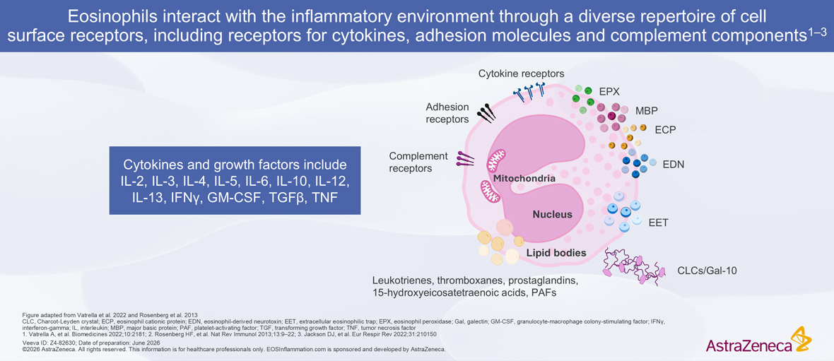
Eosinophils, which develop in the bone marrow, are differentiated effector cells that are primarily tissue-dwelling.2,5,6 Increases in recruitment to tissue, differentiation, activation, and survival of eosinophils can occur under the influence of cytokines such as IL-3, IL-5, and GM-CSF.4,5,11,12 The functional diversity of eosinophils is enabled by a repertoire of cell surface receptors, including receptors for cytokines, adhesion molecules, and complement components.4,12,13 This receptor architecture supports efficient recruitment to sites of inflammation, promotes the release of further inflammatory mediators, and can amplify activation in response to local cytokine release.4,12,13


Upon activation, eosinophils release a mixture of granules (eg EPX, MBP, ECP, and EDN) that can drive tissue dysfunction and initiate downstream inflammatory signaling.12 They also generate EETs and CLCs, which can further propagate tissue damage and sustain local immune cell activation.2


Eosinophil Inflammatory Environment Diagram Eosinophil Inflammatory Environment Diagram


Although eosinophils are present in the tissues of healthy humans, their precise physiological role remains incompletely understood.2,3 Much of what is known about eosinophil biology comes from research using in vitro models and animal studies, which provide valuable insights but do not fully clarify their roles in humans.3


Proposed roles for eosinophils span both immune and non-immune domains.14,15 Within the immune system, it has been shown that eosinophils can contribute to innate and adaptive immunity, acting as early responders to environmental stimuli while also influencing the behavior of other immune cells.14,15 Beyond immunity, eosinophils have also been proposed to play a role in reproductive homeostasis.14 Additional hypotheses point toward contributions to mammary gland development, as well as the maintenance of gastrointestinal homeostasis, where eosinophils are consistently present in the healthy gut.14

Can we live without eosinophils?

Importantly, long-term eosinophil depletion studies in humans have not been associated with significant physiological abnormalities, increased susceptibility to infection, or heightened malignancy risk.3,16 This observation has led to the hypothesis that humans possess sufficient immune redundancy, including alternative pathways and cell types capable of compensating for eosinophil loss, thereby preventing noticeable negative clinical consequences when eosinophils are depleted.3

Back to top


Eosinophilic inflammation in disease pathology

Eosinophilic inflammation in tissues is now recognized as a key contributor to the pathophysiology of several diseases, including SEA, EGPA, and HES.2,4,5,16,17


In these conditions, eosinophils become drivers of chronic inflammation, which can lead to organ dysfunction.2,4 The specific consequences vary by disease, but can include:

  • Airway remodeling, excess mucus secretion, and airway hyperresponsiveness, leading to reduced lung function, uncontrolled airway inflammation, and frequent exacerbations in SEA22,24–29
  • Organ involvement in HES and EGPA, potentially affecting the heart, lungs, skin, or gastrointestinal tract19,20,23,30

Certain eosinophil-driven diseases are associated with elevated mortality rates.31–33 This is particularly evident in systemic diseases, such as EGPA and HES, where uncontrolled eosinophilic inflammation may result in life-threatening complications, including cardiomyopathy or vasculitis.33–35


Owing to their central role in progression of these eosinophil-driven diseases, targeting eosinophils to reduce inflammation has become an established disease management strategy.7,8


Learn more about the role of eosinophils in SEA, EGPA, and HES in the dedicated educational modules

Mechanisms of eosinophil development, activation, and tissue damage

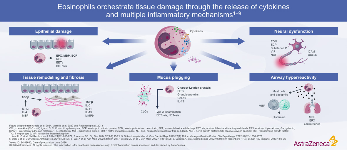
Eosinophil-Mediated Tissue Damage Diagram Eosinophil-Mediated Tissue Damage Diagram


When eosinophil progenitors in the bone marrow mature, they are released into the circulation and migrate to sites of inflammation, such as the airway tissue.13,36


Eosinophils are key effector cells within the inflammatory cascade and can contribute to tissue damage through a complex network of cellular interactions and cytokine signaling.13,36–41 Increases in recruitment to tissue, differentiation, activation, and survival of eosinophils can occur under the influence of several cytokines, including IL-3, IL-5, and GM-CSF.4,5,11


Once present in the affected tissue, eosinophils can play an important role as pathogenic cells and contribute to:

  • A perpetuated cycle of amplified inflammation, resulting in further epithelial damage and release of epithelial-derived cytokines, which can in turn activate eosinophils36,38
  • Heightened airway remodeling and hyperresponsiveness37
  • Enhanced mast cell activation and survival, resulting in additional release of inflammatory mediators, including histamines, prostaglandins, and cytokines, into the local environment36
  • IL-5 production via autocrine signaling, resulting in amplification of downstream eosinophil effects41
  • T-cell proliferation, differentiation, and apoptosis36

Through these interconnected pathways, eosinophils can become key drivers of tissue damage and dysfunction in disease.13,36–39,41 Eosinophil levels
have been found to be raised across a heterogenous range of eosinophil-associated diseases, including SEA, EGPA, HES, IBD, atopic dermatitis, and CRSwNP.1,42–44


 

Downstream effects of eosinophilic inflammation may include:

Epithelial damage


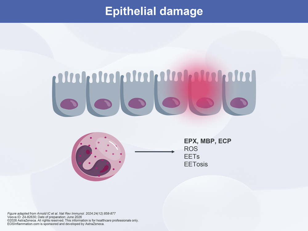
Release of eosinophil-derived proteins such as EPX, MBP, and ECP have been shown to directly injure the epithelium.1 These toxic mediators, along with release of ROS via EETosis, can result in compromised epithelial barrier integrity and persistent release of inflammatory mediators in diseases such as SEA, EGPA, and HES.1

Epithelial Damage Diagram Epithelial Damage Diagram

Tissue remodeling and fibrosis


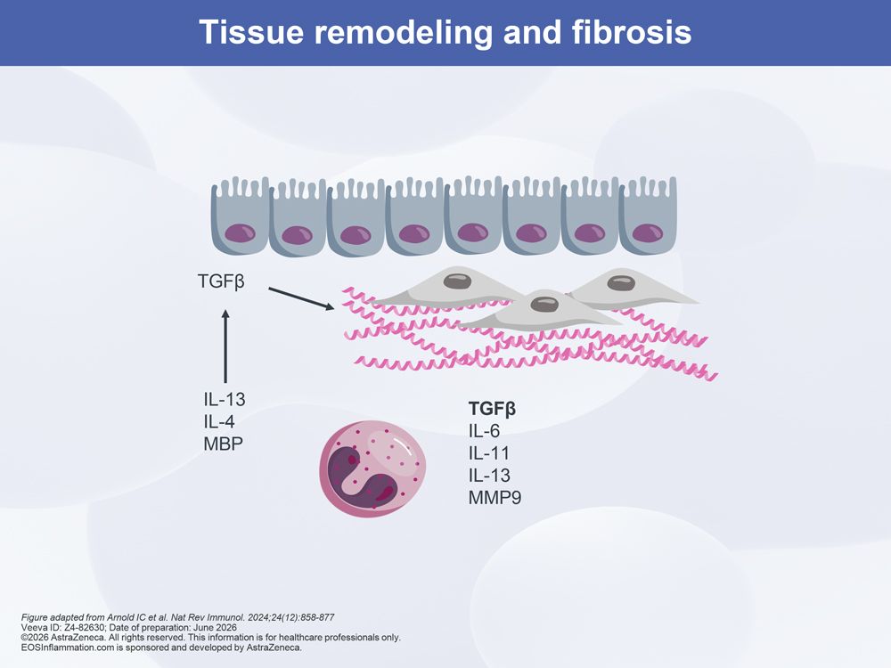
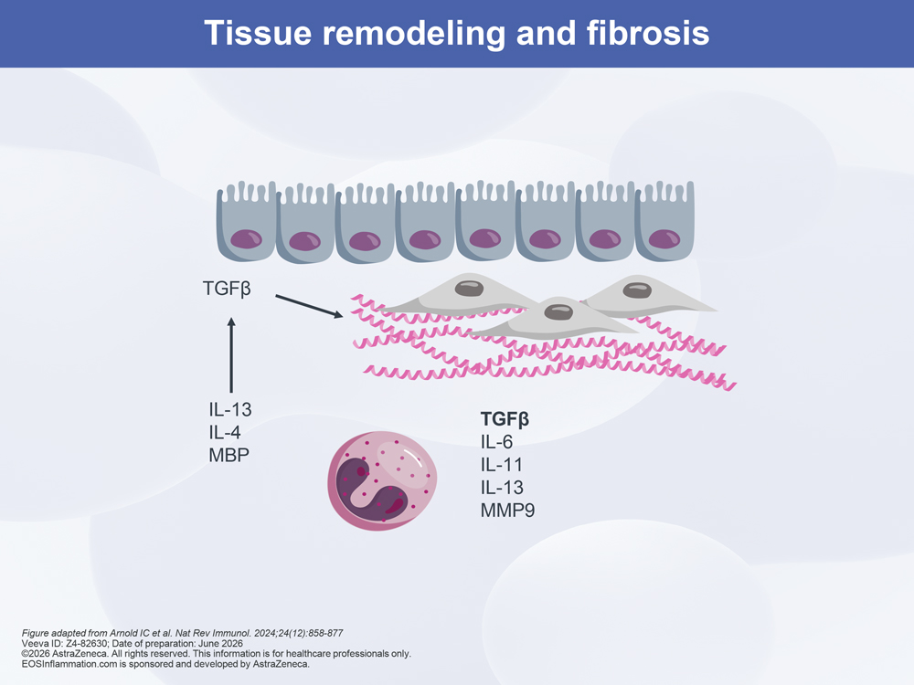
Chronic eosinophilic inflammation can promote structural changes.1 The release of cytokines IL-13 and IL-4, as well as MBP, by eosinophils can drive fibroblast proliferation and extracellular matrix deposition via TGF-β, IL-6, IL-11, IL-13, and matrix metalloproteinases (eg MMP9). This can result in long-term tissue remodeling and even fibrosis in SEA, HES, and EGPA.1,18,44 The resulting clinical consequences of eosinophil-associated remodeling and eosinophil-driven fibrosis are often severe, with lasting impacts on patient quality of life, such as endomyocardial fibrosis and heart failure in HES, and airway remodeling and irreversible airway obstruction in SEA.18

Tissue Remodeling And Fibrosis Diagram Tissue Remodeling And Fibrosis Diagram

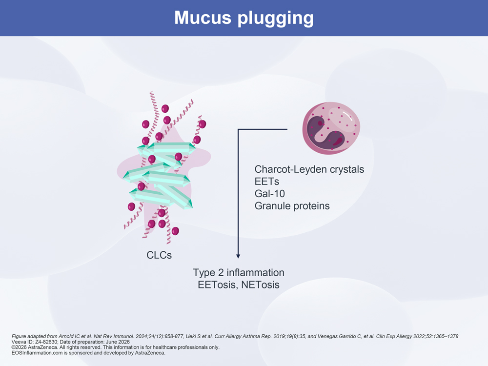
Mucus plugging


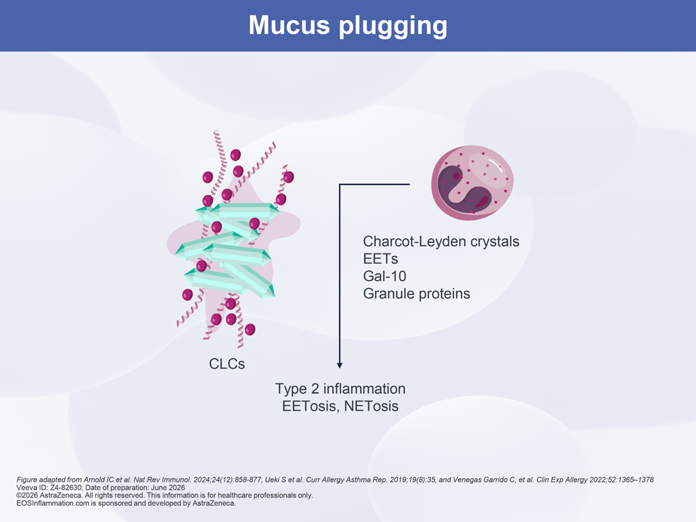
Activated eosinophils can undergo EETosis, resulting in the release of granule proteins, CLCs, as well as EVs and ETs.1,21,45 These structures have been shown to increase the viscosity of mucus and promote plugging, which can cause airflow obstruction.1,21,45 Furthermore, release of leukotrienes and cytokines, such as IL-13, can promote mucus hypersecretion.1 Together, these mechanisms perpetuate type 2 inflammation and increase the probability of mucus plugging in asthma.18,21,45 Importantly, mucus within the airways is increasingly being recognized as a critical driver of disease symptoms through obstruction of the airway lumen and exacerbations.21,46,47

MUCUS PLUGGING DIAGRAM MUCUS PLUGGING DIAGRAM

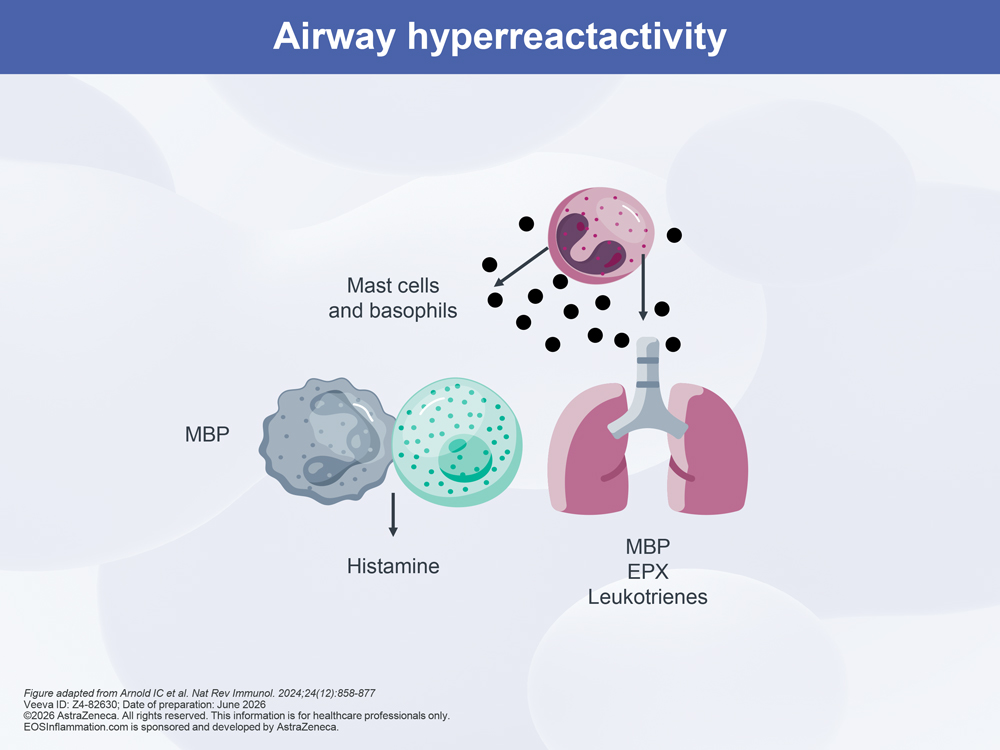
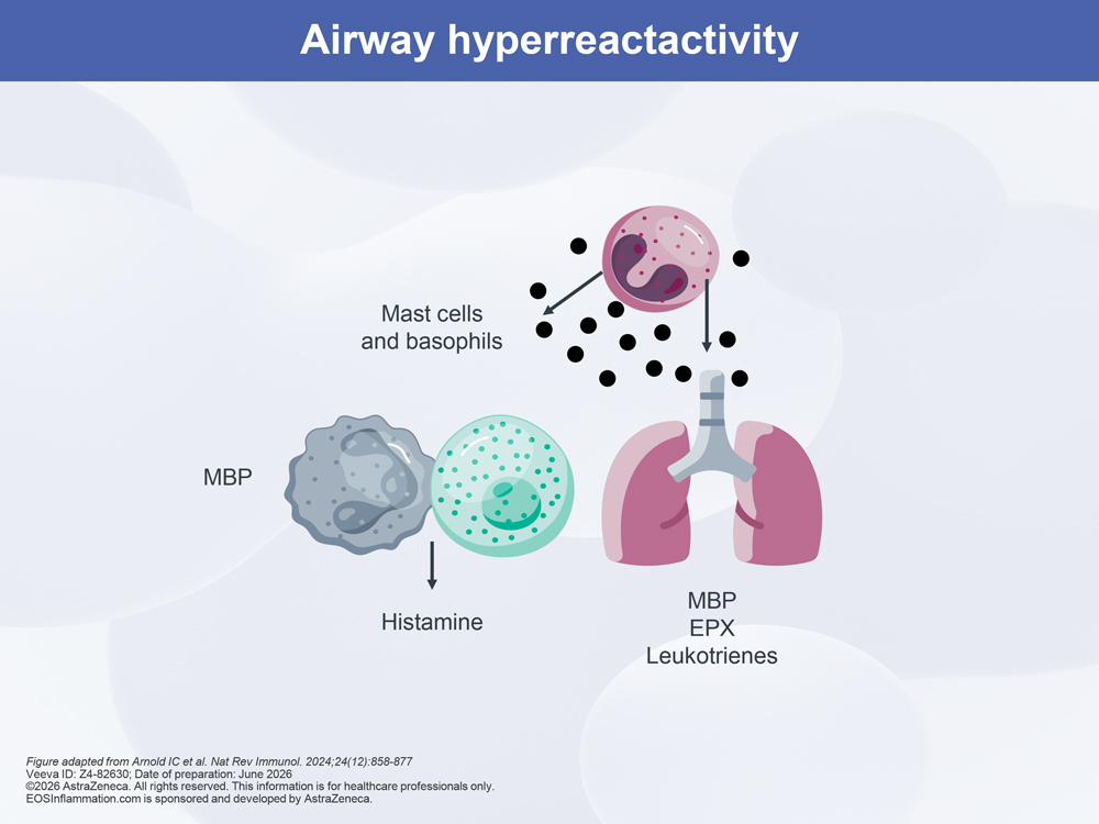
Airway hyperreactivity


MBP, EPX, leukotrienes, and cytokines such as IL-13, released from eosinophils, are known to contribute to airway hyperresponsiveness.1 The release of mediators such as MBP and EPX has been shown to damage lung structural cells and promote airway hyperreactivity, which manifests as hallmark features of asthma such as bronchospasm, wheezing/coughing, airflow limitation, and dyspnea.1,43

Airway Hyperreactivity Diagram Airway Hyperreactivity Diagram

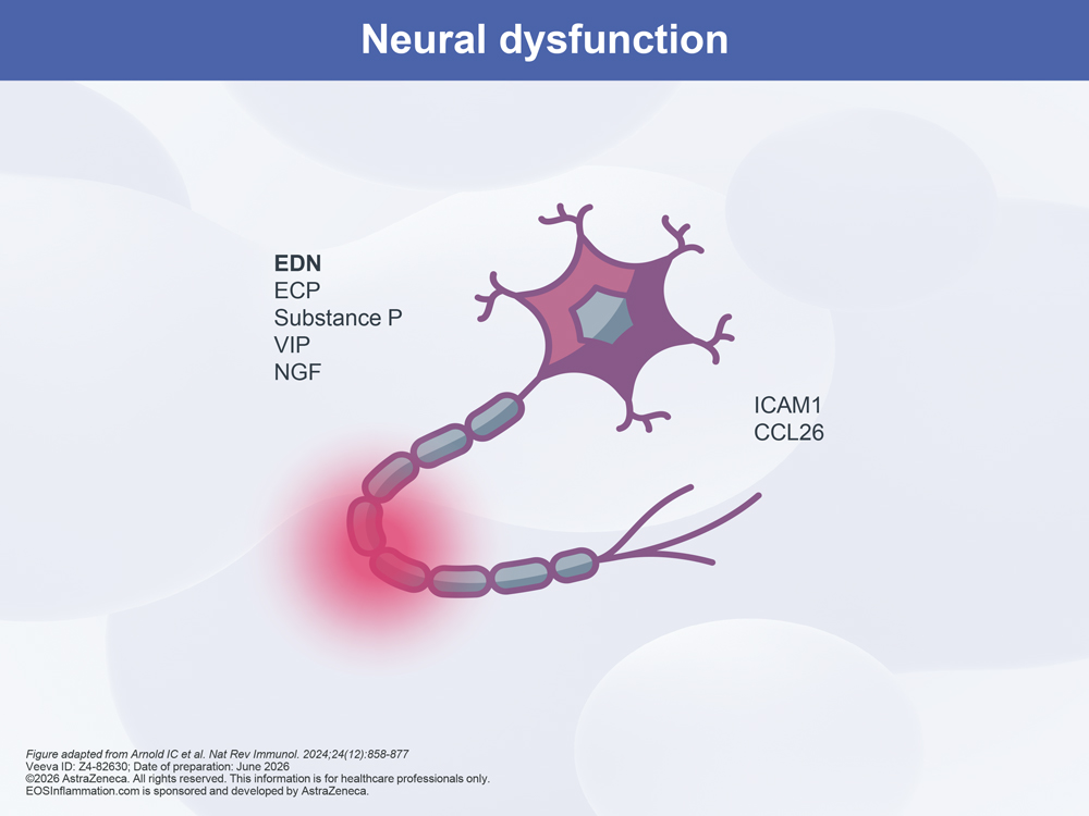
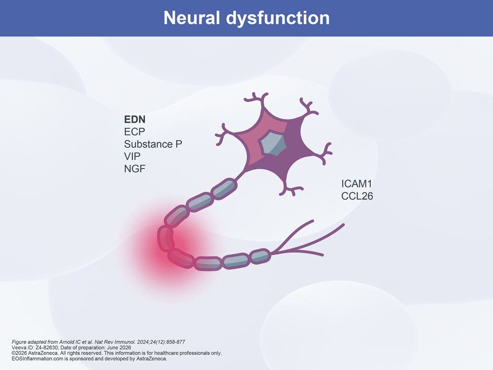
Neural dysfunction


Eosinophils can impact neural signaling via the release of mediators such as EDN, ECP, substance P, VIP, and NGF.1,42 In certain diseases, eosinophils can accumulate in close proximity to neural fibers and ganglia and have been hypothesized to exert both neuroinflammatory and neurotoxic effects.1,42

Neural Dysfunction Diagram Neural Dysfunction Diagram
  1. Epithelial-damage
  2. Tissue-remodeling
  3. Mucus-plugging
  4. Airway-hyperreactivity
  5. Neural-dysfunction


Back to top

Corticosteroids as a management approach for eosinophilic inflammation

Management of eosinophil-driven diseases such as SEA, EGPA, and HES has historically relied on the use of corticosteroids, both local and systemic, to reduce the effects of inflammation.9 This is reflected in their inclusion in international guidelines for disease management and treatment.9,48–50


While both OCS and ICS can impact the inflammatory cascade, ICS are likely to act on the epithelium, reducing eosinophil recruitment, while OCS are likely to deplete the reservoir of circulating eosinophils.51


It is now widely recognized that corticosteroids are associated with adverse effects and corticoresistance, and that poor disease control is often reported despite their use.9 Reporting of adverse events is particularly associated with the use of systemic corticosteroids at high dose, for repeated cycles, or for a prolonged period of time.9


For further information on the role of corticosteroids in the management of eosinophil-driven diseases, visit the disease-specific educational modules

Back to top

Eosinophils as prognostic markers supporting disease management

Some researchers suggest that eosinophils play a central role not only in driving inflammation but also in predicting disease course.4,10,52–54 These cells are considered a potential prognostic marker in several eosinophil-driven diseases, and have been explored for their ability to support a predict and prevent approach to disease management.4,10,52–54

Back to top

CLC, Charcot-Leyden crystal; CRSwNP, chronic rhinosinusitis with nasal polyps; ECP, eosinophil cationic protein; EDN, eosinophil-derived neurotoxin; EET, extracellular eosinophilic trap; EETosis, eosinophil extracellular trap cell death; EGPA, eosinophilic granulomatosis with polyangiitis; EOS, eosinophil; EPX, eosinophil peroxidase; ET, eosinophilic trap; EV, extracellular vesicle; GM-CSF, granulocyte-macrophage colony-stimulating factor; HES, hypereosinophilic syndrome; IBD, inflammatory bowel disease; ICS, inhaled corticosteroid(s); IL, interleukin; MBP, major basic protein; MMP, matrix metalloproteinase; NGF, nerve growth factor; OCS, oral corticosteroid(s); ROS, reactive oxygen species; SEA, severe eosinophilic asthma; TGF-β, transforming growth factor beta; VIP, vasoactive intestinal peptide

  1. Arnold IC, Munitz A. Nat Rev Immunol 2024;24:858–877
  2. Jacobsen EA, et al. Annu Rev Immunol 2021;39:719–757
  3. Jackson DJ, Pavord ID. Eur Respir J 2023;61:2201217
  4. Quirce S, et al. Front Immunol 2023;14:1192284
  5. Klion AD, et al. Annu Rev Pathol 2020;15:179–209
  6. Weller PF, Spencer LA. Nat Rev Immunol 2017;17:746–760
  7. Fulkerson PC, Rothenberg ME. Nat Rev Drug Discov 2013;12:117–129
  8. Jackson DJ, et al. Eur Respir Rev 2022;31:210150
  9. Del Pozo V, et al. Front Immunol 2023;14:1310211
  10. Couillard S, et al. J Allergy Clin Immunol Pr 2023;11:704–712
  11. Park YM, Bochner BS. Allergy Asthma Immunol Res 2010;2:87–101
  12. Vatrella A, et al. Biomedicines 2022;10:2181
  13. Rosenberg HF, et al. Nat Rev Immunol 2013;13:9–22
  14. Marichal T, et al. Front Med (Lausanne) 2017;4:101
  15. Ramirez GA, et al. Biomed Res Int 2018;2018:9095275
  16. Gleich GJ, et al. Allergy 2013;68:829–835
  17. Rothenberg ME, Hogan SP. Annu Rev Immunol 2006;24:147–174
  18. Aceves SS. Dig Dis 2014;32:15–21
  19. Chakraborty RK, Aeddula NR. Eosinophilic granulomatosis with polyangiitis (Churg-Strauss syndrome). Treasure Island, FL, USA:
StatPearls Publishing, 2023
  20. Khoury P, et al. Nat Rev Rheumatol 2014;10:474–483
  21. Venegas Garrido C, et al. Clin Exp Allergy 2022;52:1365–1378
  22. Hussain M, Liu G. Cells 2024;13:384
  23. Fagni F, et al. Front Med (Lausanne) 2021;8:627776
  24. McBrien CN, Menzies-Gow A. Front Med (Lausanne) 2017;4:93
  25. Hogan SP, et al. Clin Exp Allergy 2008;38:709–750
  26. Siddiqui S, et al. J Allergy Clin Immunol 2023;152:841–857
  27. Chan R, et al. J Allergy Clin Immunol Pr 2023;11:1459–1462
  28. Dunican EM, et al. J Clin Invest 2018;128:997–1009
  29. Tang M, et al. Am J Respir Crit Care Med 2022;205:1036–1045
  30. Kovacs N, et al. J Allergy Clin Immunol Pr 2020;8:3209–3212
  31. Hospers JJ, et al. Epidemiology 2000;11:261–268
  32. Hartog NL, Beigelman A. J Allergy Clin Immunol 2018;141:AB276
  33. Taniguchi M, et al. Allergol Int 2007;56:97–103
  34. Trivioli G, et al. Rheumatol Oxf 2020;59(Suppl. 3):iii84–iii94
  35. Akuthota P, Weller PF. Immunol Allergy Clin North Am 2015;35:403–411
  36. Trivedi SG, Lloyd CM. Cell Mol Life Sci 2007;64:1269–1289
  37. Patterson MF, et al. J Asthma Allergy 2015;8:125–134
  38. Lambrecht BN, Hammad H. Nat Immunol 2015;16:45–56
  39. Denburg JA, Keith PK. Chest 2008;134:1037–1043
  40. Gandhi NA, et al. Nat Rev Drug Discov 2016;15:35–50
  41. AbuJabal R, et al. Clin Exp Allergy 2024;54:538–549
  42. Cioclu MC, et al. J Clin Med 2022;11:5595
  43. Wei X, et al. Ann Med 2022;54:11–21
  44. Srikantharajah M, et al. Curr Cardiol Rep 2025;27:109
  45. Ueki S, et al. Curr Allergy Asthma Rep 2019;19:35
  46. Aegerter H, Lambrecht BN. Annu Rev Pathol Mech Dis 2023;18:387–409
  47. Ueki S, et al. J Allergy Clin Immunol Pr 2026;14:575–582
  48. Global Initiative for Asthma (GINA). Global strategy for asthma management and prevention. 2026. Available from: https://ginasthma.org/2026-gina-strategy-report/ (Accessed June 11, 2026)
  49. Hellmich B, et al. Ann Rheum Dis 2024;83:30–47
  50. Shomali W, Gotlib J. Am J Hematol 2024;99:946–968
  51. Pavord ID. Curr Opin Pulm Med 2019;25:51–58
  52. Khoury P, et al. Front Med (Lausanne) 2017;4:240
  53. Lommatzsch M, et al. Lancet Respir Med 2025;13:943–950
  54. Matucci A, et al. Biomedicines 2022;10:2688
AZ-logo

EOSInflammation.com has been
developed by AstraZeneca.
For healthcare professionals only.
© 2026 AstraZeneca

  • Privacy Notice
  • Cookie Notice
  • Legal Statement
  • Contact Us

Veeva ID: Z4-82630
Date of preparation: June 2026